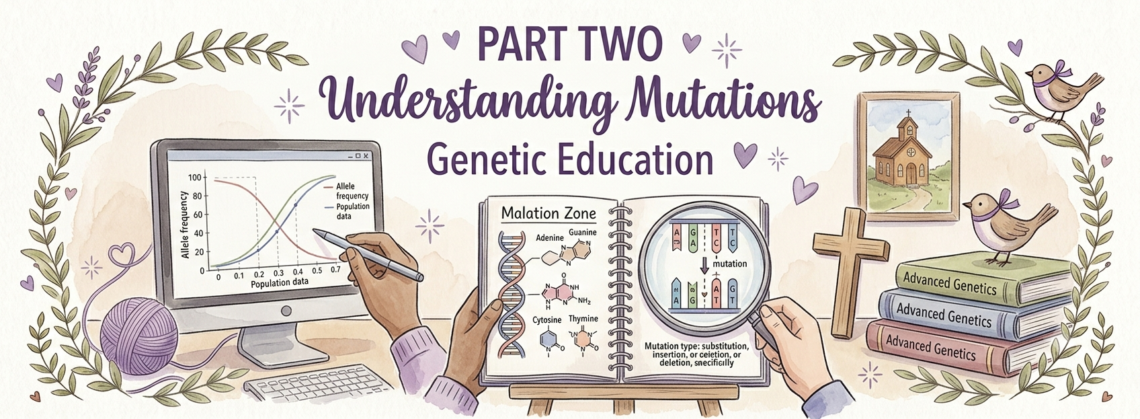
In this Part Two is where I will break down how the mutations work and how to understand your mutation!
Note: anything highlighted yellow is either something important or is the start of a new topic, if highlighted in pink indicates that that specific thing represents my mutations as well.
THE FUNDAMENTALS OF DNA AND GENES
What Is DNA
Deoxyribonucleic acid (DNA) is the molecule that stores the genetic instructions for building, running, and maintaining every living organism. It is present in virtually every cell of your body (with rare exceptions like red blood cells, which lose their nucleus as they mature).
Structure of DNA:
DNA is a double helix — two long chains wound around each other like a twisted ladder. Each chain is made of repeating units called nucleotides. Each nucleotide consists of:
- A sugar molecule (deoxyribose)
- A phosphate group
- One of four nitrogenous bases:
- Adenine (A)
- Thymine (T)
- Guanine (G)
- Cytosine (C)
- The two chains of the double helix are held together by hydrogen bonds between complementary base pairs:
A always pairs with T (two hydrogen bonds)
- G always pairs with C (three hydrogen bonds)
This complementary base pairing is fundamental to every process that uses DNA — replication, transcription, and repair.
The human genome:
- Contains approximately 3.2 billion base pairs of DNA
- Is organized into 23 pairs of chromosomes (46 total in most cells) — 22 pairs of autosomes plus one pair of sex chromosomes (XX in females, XY in males)
- Contains approximately 20,000–25,000 protein-coding genes (only about 1.5% of total DNA)
- The remaining ~98.5% includes regulatory sequences, introns (non-coding gene regions), repetitive elements, and regions of currently unknown function
From DNA to Protein — The Central Dogma
The flow of genetic information follows the Central Dogma of Molecular Biology:
DNA → (Transcription) → mRNA → (Translation) → Protein
Step 1 — Transcription:
The DNA sequence of a gene is copied into a single-stranded messenger RNA (mRNA) molecule by an enzyme called RNA polymerase. The mRNA is a complementary copy of one strand of the DNA double helix (using RNA nucleotides: A, U, G, C — note Uracil replaces Thymine in RNA).
During transcription in eukaryotes (organisms with a nucleus, including humans), the initial RNA copy (pre-mRNA) includes both exons (protein-coding segments) and introns (non-coding intervening sequences). The introns are then removed and the exons are spliced together in a process called RNA splicing. The mature mRNA contains only the exon sequences.
Step 2 — Translation:
The mRNA travels from the nucleus to the cytoplasm, where molecular machines called ribosomes read the mRNA sequence and build a protein. Each group of three consecutive nucleotides (a codon) specifies one amino acid. This triplet code is called the genetic code.
There are 64 possible codons (4³ = 64) but only 20 standard amino acids — so the genetic code is redundant (multiple codons can specify the same amino acid). There are also three stop codons (UAA, UAG, UGA) that signal the ribosome to end translation.
Step 3 — Post-translational modification:
After the protein chain is assembled, it typically undergoes various modifications (folding, cleavage, addition of chemical groups, assembly with other subunits) before becoming a fully functional protein.
MUTATIONS — A COMPREHENSIVE CLASSIFICATION
- A mutation is any change in the DNA sequence compared to a reference sequence. In genetics, mutations are classified in many overlapping ways depending on what aspect you’re describing.
Classification by SIZE/SCALE
- Point Mutations (Single Nucleotide Changes)
- A change affecting only ONE nucleotide.
Subtypes:
A) Substitution (Single Nucleotide Variant, SNV):
- One nucleotide is replaced by another. This is the most common type of mutation.
Example: …AATGGT… → …AATCGT… (G replaced by C at position 4) - Further classified by the type of substitution
- Transition: Substitution between same-class nucleotides (purine↔purine: A↔G; or pyrimidine↔pyrimidine: C↔T). More common because the bases are chemically similar.
- Transversion: Substitution between different-class nucleotides (purine↔pyrimidine: A/G↔C/T). Less common but more likely to cause amino acid changes.
B) Single Nucleotide Insertion:
- One nucleotide is added to the sequence where there wasn’t one before.
Always causes a frameshift (unless it occurs in a complete codon context)
C) Single Nucleotide Deletion: - One nucleotide is removed from the sequence.
This is exactly where My maternal RYR1 mutation (c.12567del) is — one nucleotide deleted, causing a frameshift - Always causes a frameshift
2. Small Insertions and Deletions (Indels)
Changes of 2 to approximately 50 nucleotides.
In-frame indels (divisible by 3):
- Delete or insert complete codons
- Reading frame is preserved
- Net effect: missing or extra amino acids in the protein
- MY PLEC mutation (c.6271_6282del — 12 nucleotide deletion = 4 codons) is this type
- Can range from mild to severe depending on which amino acids are lost/gained
Frameshift indels (NOT divisible by 3):
- Shift the reading frame from the point of change onward
- All codons from that point forward are scrambled
- Nearly always result in a premature stop codon downstream
- Nearly always trigger nonsense-mediated decay
- Nearly always result in no functional protein
- MY maternal RYR1 mutation is this type (1 nucleotide deleted = not divisible by 3 = frameshift)
3. Copy Number Variants (CNVs)
Larger scale changes involving amplification or loss of segments of DNA.
Types:
- Deletion CNV: A segment of DNA (anywhere from a few hundred base pairs to millions of base pairs) is missing
- Duplication CNV: A segment of DNA is present in extra copies (could be tandem duplications, in which the extra copy sits adjacent to the original, or dispersed duplications)
- Detected by: Array comparative genomic hybridization (aCGH), chromosomal microarray, MLPA (Multiplex Ligation-dependent Probe Amplification)
- The Invitae test that diagnosed MY also performed “deletion/duplication testing” — this would have detected CNVs in addition to point mutations and small indels
4. Structural Variants (SVs)
Changes in the architecture of the genome beyond simple copy number changes.
Types:
- Inversions: A segment of DNA is flipped in orientation (the same sequence is present but runs in reverse)
- Translocations: A segment of DNA moves from one chromosomal location to another (can be between chromosomes — interchromosomal translocation — or within the same chromosome — intrachromosomal translocation)
- Ring chromosomes: The two ends of a chromosome fuse to form a circular chromosome
- Isochromosomes: A chromosome that has two copies of one arm and no copy of the other
5. Chromosomal Aneuploidies
Changes in the NUMBER of entire chromosomes.
Types:
- Monosomy: Only one copy of a chromosome instead of the normal two (e.g., Turner syndrome = 45,X — only one sex chromosome)
- Trisomy: Three copies of a chromosome instead of two (e.g., Down syndrome = trisomy 21; Edward syndrome = trisomy 18; Patau syndrome = trisomy 13)
- Polyploidy: Additional complete sets of chromosomes (triploid = 69 chromosomes; tetraploid = 92 chromosomes) — generally lethal in humans except in some cancer contexts
Classification by FUNCTIONAL CONSEQUENCE
This classification system describes what the mutation DOES to the protein or gene function, regardless of its size.
1. Silent Mutations (Synonymous Mutations)
A nucleotide change that does NOT change the amino acid.
- Possible because the genetic code is redundant — multiple codons specify the same amino acid
- Example: Both GCU and GCC code for Alanine; changing the third position C→U produces the same amino acid
- Not truly silent: Despite the name, these can affect: mRNA stability, translation efficiency, splicing regulatory sequences, and RNA secondary structure — so “silent” mutations can sometimes be pathogenic despite no amino acid change
- Population frequency: Common and generally benign; comprise a large portion of normal human genetic variation
2. Missense Mutations
A nucleotide change that results in a DIFFERENT amino acid being incorporated at that position.
- The most common type of pathogenic point mutation
- Effect severity depends on: which amino acid is changed, what it’s changed TO, and where in the protein it sits
- MY three paternal RYR1 variants (p.Arg3366His, p.Tyr3933Cys, p.Ile1571Val) are all missense mutations
Subtypes of missense mutations by severity:
Conservative missense:
- Replacement with an amino acid of similar chemical character
- Example: Ile→Val (both nonpolar, hydrophobic, similar size) — MY p.Ile1571Val is the most conservative
- Often tolerated without significant functional impact
- But: even conservative changes at critical sites can be pathogenic (as with MY p.Ile1571Val which is classified VUS despite the conservative chemistry, because the isoleucine residue is highly conserved)
Non-conservative missense:
- Replacement with an amino acid of very different chemical character
- Example: Tyr→Cys (aromatic, hydrogen-bonding → small, sulfhydryl; large size difference, different reactivity) — MY p.Tyr3933Cys is a non-conservative missense
- Much more likely to disrupt protein function
- More likely to be pathogenic
Semi-conservative missense:
- In between — some chemical similarity lost but not completely different
- Example: Arg→His (both positively charged, but different size and charge strength) — MY p.Arg3366His falls in this category
3. Nonsense Mutations (Stop-Gain Mutations)
A nucleotide change that creates a premature stop codon (UAA, UAG, or UGA) within the protein-coding sequence.
- The ribosome stops translation prematurely at the new stop codon
- Results in a truncated protein — missing everything C-terminal to the stop codon
- If the premature stop codon is more than ~55 nucleotides upstream of the last exon-exon junction, nonsense-mediated decay (NMD) typically degrades the mRNA before the truncated protein is made
- Generally loss-of-function — the protein either isn’t made or lacks critical domains
- Distinguished from MY maternal mutation (which is technically classified as a frameshift with premature stop) — a pure nonsense mutation changes a sense codon directly to a stop codon, while MY mutation creates a stop codon indirectly through the frameshift reading frame
4. Frameshift Mutations
Insertions or deletions of a number of nucleotides NOT divisible by 3.
- Shifts the reading frame from the point of mutation onward
- All codons downstream are scrambled
- Almost always generates a premature stop codon somewhere downstream
- Almost always triggers NMD → no protein from that allele
- MY maternal RYR1 mutation is a frameshift mutation — specifically a frameshift deletion (1 nucleotide deleted)
- Generally considered the most severe class of mutation along with large deletions and nonsense mutations
5. Splice Site Mutations
Mutations affecting the sequences that signal where exons and introns begin and end.
Background on splicing:
- Before mRNA can be translated into protein, the initial RNA transcript (pre-mRNA) must have its introns removed and exons joined together. This process (RNA splicing) is controlled by:
- Splice donor site: The sequence at the beginning of an intron (exon-intron boundary), typically beginning with GT in the DNA (GU in RNA)
- Splice acceptor site: The sequence at the end of an intron (intron-exon boundary), typically ending with AG in the DNA
- Branch point sequence: A conserved sequence within the intron that helps identify the correct splice site
- Splicing enhancers and silencers: Regulatory sequences within exons and introns that help or hinder recognition of nearby splice sites
Effects of splice site mutations:
- Exon skipping: The exon adjacent to the mutated splice site is not recognized and is excluded from the mRNA (as though it doesn’t exist)
- Intron retention: The intron adjacent to the mutated site is not removed — stays in the mRNA, typically disrupting the reading frame
- Cryptic splice site activation: The normal splice site is disrupted, and the splicing machinery uses an alternative (cryptic) sequence nearby — can create aberrant mRNA with insertions or deletions of various sizes
- All of these typically result in loss-of-function — the protein is either absent (NMD) or made in an aberrant form
6. In-Frame Insertions and Deletions
Insertions or deletions of a number of nucleotides exactly divisible by 3.
- Reading frame is preserved
- Result in the addition or removal of complete amino acids
- MY PLEC mutation (12-nucleotide deletion = 4 amino acids removed) is an in-frame deletion
- Effect ranges from negligible (if removed/added amino acids are in non-critical regions) to severe (if in critical domains)
- Generally NOT subject to NMD (protein IS made, just shorter or longer)
7. Repeat Expansion Mutations
A specific type of mutation where a short tandemly repeated DNA sequence expands in copy number beyond the normal range.
- Normal individuals have a certain number of repeats (e.g., 10–35 copies of a trinucleotide repeat)
- Affected individuals have an expanded number (e.g., hundreds or thousands of copies)
Examples of diseases caused by repeat expansions:
- Huntington’s disease: CAG repeat expansion in the HTT gene (>36 repeats causes disease; >60 repeats causes juvenile onset)
- Fragile X syndrome: CGG repeat expansion in the FMR1 gene (>200 repeats causes intellectual disability)
- Myotonic dystrophy type 1: CTG repeat expansion in the DMPK gene — notably a muscular dystrophy
- Myotonic dystrophy type 2: CCTG repeat expansion in CNBP gene
- Friedreich’s ataxia: GAA repeat expansion in the FXN gene
- ALS/FTD (some forms): GGGGCC repeat expansion in C9orf72 gene
Repeat expansions can act through various mechanisms: loss of the normal gene product, production of a toxic expanded RNA, production of a toxic expanded protein, or all three simultaneously
8. Regulatory Mutations (Non-Coding Mutations)
Mutations in DNA sequences that don’t code for protein but regulate gene expression
Types of regulatory regions:
- Promoter: The sequence immediately upstream (before) a gene where RNA polymerase binds to initiate transcription. Mutations here can reduce or eliminate gene expression.
- Enhancer: Sequences (often far from the gene, sometimes in introns or even millions of base pairs away) that increase gene transcription when bound by transcription factors.
- Silencer: Sequences that decrease gene transcription.
- Insulator: Sequences that block the influence of enhancers or silencers on adjacent genes.
- 5′ UTR (Untranslated Region): The sequence in the mRNA between the start of transcription and the start codon; mutations here can affect mRNA stability and translation efficiency.
- 3′ UTR: The sequence after the stop codon; contains signals for mRNA stability, localization, and translation regulation including microRNA binding sites.
- Splice regulatory sequences (within exons and introns): Exonic splicing enhancers (ESEs), exonic splicing silencers (ESSes), intronic splicing enhancers (ISEs), intronic splicing silencers (ISSes) — all can be mutated to cause aberrant splicing even when the canonical splice sites themselves are unchanged.
Why regulatory mutations matter:
Standard exome sequencing (which reads only the protein-coding exon sequences) MISSES regulatory mutations entirely. Only whole-genome sequencing captures the full regulatory landscape. This means patients with a clinical presentation strongly suggesting a genetic disease but with negative exome sequencing may have mutations in regulatory regions that went undetected.
9. Epigenetic Mutations
Changes in gene expression that do NOT alter the DNA sequence itself but alter how the DNA is read.
Types:
- DNA methylation changes: Methyl groups (-CH₃) are added to cytosine bases (typically at CpG dinucleotides). Methylated genes are generally silenced; unmethylated genes are generally active. Abnormal methylation can silence tumor suppressor genes (cancer) or activate normally silent sequences.
- Histone modification changes: DNA is wrapped around proteins called histones. Chemical modifications to histones (acetylation, methylation, phosphorylation, ubiquitination) alter how tightly or loosely the DNA is packaged, affecting which genes can be transcribed.
- Imprinting disorders: Some genes are normally expressed from only one parental copy (the other is epigenetically silenced). Mutations in imprinting control regions cause both copies to be silenced (or both expressed), leading to disease. Examples: Prader-Willi syndrome and Angelman syndrome (both involve chromosome 15q11-q13 but different parental copies).
Classification by INHERITANCE PATTERN
1. Autosomal Dominant
- ONE mutated copy of a gene is sufficient to cause disease
- The mutated gene is on one of the autosomes (chromosomes 1-22, not sex chromosomes)
- An affected parent has a 50% chance of passing the mutation to each child regardless of sex
- Examples: Huntington’s disease, BRCA1/2 hereditary breast/ovarian cancer, Marfan syndrome, Neurofibromatosis type 1, most forms of CCD from RYR1
Mechanisms of dominance:
- Haploinsufficiency: One functional copy of the gene doesn’t produce enough protein for normal function (50% of normal protein is insufficient)
- Dominant negative: The mutant protein is made and actively interferes with the function of the normal protein from the other allele (especially relevant for proteins that form multimers/complexes — like RYR1’s homotetramer)
- Gain of toxic function: The mutant protein acquires a new harmful activity that the normal protein doesn’t have (e.g., Huntington’s disease — the expanded polyglutamine protein aggregates and is toxic)
2. Autosomal Recessive
- TWO mutated copies of a gene are required to cause disease
- Both copies on autosomes must be affected
- Parents are typically carriers — they have one normal and one mutated copy and are generally unaffected
- Two carrier parents have a 25% chance of an affected child (inherits mutation from both parents), 50% chance of a carrier child (inherits one mutation), and 25% chance of a completely unaffected child (inherits neither mutation) with each pregnancy
Two ways to have two mutated copies:
- Homozygous: Both copies have the SAME mutation (inherited one copy from each carrier parent, or both mutations arose independently but happen to be identical)
- Compound heterozygous: The two copies have DIFFERENT mutations — one from each parent. This is MY situation — one maternal null allele plus one paternal three-VUS allele.
- Examples: Cystic fibrosis, sickle cell disease, phenylketonuria (PKU), spinal muscular atrophy (SMA), most severe RYR1 myopathies (including MY), many lysosomal storage diseases
3. X-Linked Recessive
- The mutated gene is on the X chromosome
- Males (XY) have only ONE X chromosome, so one mutated copy causes disease (they have no second X copy to compensate)
- Females (XX) typically need TWO mutated X copies to be affected (they usually have one normal X to compensate); females with one mutated copy are carriers and are usually unaffected (though carrier females can sometimes have mild symptoms)
- Pattern: typically skips generations in families (grandfather → carrier daughters → affected grandsons)
- Examples: Duchenne Muscular Dystrophy (DMD), Becker Muscular Dystrophy (BMD), hemophilia A and B, color blindness, Fabry disease (though this can also affect carrier females), Hunter syndrome (MPS II)
4. X-Linked Dominant
- The mutated gene is on the X chromosome AND one mutated copy is sufficient to cause disease
- Both males and females can be affected, though often more severely in one sex
- Affected fathers pass the mutation to ALL daughters (who have XX; they’ll receive the father’s X) but NONE of their sons (who receive the father’s Y)
- Affected mothers have a 50% chance of passing it to both sons and daughters
Examples: Rett syndrome (affects females almost exclusively — males with MECP2 mutations rarely survive to birth), Incontinentia pigmenti, some forms of CHARGE syndrome
5. Y-Linked (Holandric)
- Mutated gene is on the Y chromosome
- Affects only males
- Passes from father to ALL sons (no daughters, who receive the X from father)
- Very few diseases — the Y chromosome has very few genes
- Example: Some forms of azoospermia (absence of sperm) due to AZF region deletions
6. Mitochondrial Inheritance
- Mutations in mitochondrial DNA (mtDNA) — the small circular genome inside mitochondria, separate from the nuclear genome
- Mitochondria are inherited almost exclusively from the MOTHER (the egg contributes virtually all mitochondria; sperm mitochondria are typically degraded after fertilization)
- Therefore: an affected mother passes mitochondrial mutations to ALL of her children (sons and daughters); an affected father does NOT pass mitochondrial mutations to any children
- A unique feature: heteroplasmy — a cell may contain a mixture of normal and mutated mitochondria. The proportion of mutated mitochondria (heteroplasmy level) can vary between tissues and individuals, and disease severity correlates with the proportion of mutated mitochondria.
- Examples: MELAS (Mitochondrial Encephalomyopathy, Lactic Acidosis, and Stroke-like episodes), MERRF (Myoclonic Epilepsy with Ragged-Red Fibers), Leber Hereditary Optic Neuropathy (LHON), Leigh syndrome (some forms)
- Relevant to ME: My RYR1 disease affects mitochondrial function SECONDARILY (through disrupted calcium signaling to mitochondria) — but My mutations are in NUCLEAR DNA (chromosome 19), not in mitochondrial DNA. This is an important distinction.
7. Digenic Inheritance
- Two mutations in TWO DIFFERENT GENES are BOTH required to cause disease
- Neither mutation alone is sufficient
- A relatively recently recognized inheritance pattern
Example: Bardet-Biedl syndrome can require mutations in two different BBS genes simultaneously; some forms of retinitis pigmentosa are digenic
8. Oligogenic Inheritance
- Mutations in several (a small number of) genes together determine disease risk and severity
- Increasingly recognized as underlying the variable expressivity seen in many “single-gene” diseases — modifier genes in the background influence how a primary disease gene mutation manifests
9. Polygenic and Multifactorial Inheritance
- Many genes EACH contributing small effects, combined with environmental factors
- No single gene variant is necessary or sufficient
- Examples: Most common diseases (heart disease, type 2 diabetes, hypertension, many psychiatric conditions, common cancers)
- Studied using genome-wide association studies (GWAS) which identify genomic regions associated with disease risk across large populations
Classification by ORIGIN
De Novo Mutations
- Mutations that arise NEW in a child — not inherited from either parent
- The child’s DNA has the mutation, but neither parent’s DNA has it (in their germline)
- Arise from errors in DNA replication during gametogenesis (formation of eggs or sperm) or in the earliest cell divisions after fertilization
- Rate: Approximately 30–70 new de novo mutations per generation in humans (varies by parental age — older parents, especially older fathers, have higher de novo mutation rates)
- Important in dominant diseases: explain cases where affected children are born to unaffected parents (neither parent has the mutation, but it arose de novo in the child)
- MY novel pathogenic RYR1 variant (c.12567del) may be a de novo mutation in one of My parents — most likely arising in My mother (since it’s the maternal allele), possibly as a de novo event in My grandmother or further back in the maternal line, given that it’s never been seen in any population database
Inherited Mutations
Passed from parent to child - Present in the parent’s germline DNA
- The three paternal VUS variants are clearly inherited — they came from My father’s germline
Somatic Mutations
- Arise in non-germline body (somatic) cells after conception — AFTER the egg was fertilized
- Present in some cells of the body but NOT in the germline (eggs/sperm)
- Therefore NOT heritable — cannot be passed to children
- Accumulate throughout life (aging, environmental damage)
- Critical in cancer: most cancers are caused by somatic mutations in oncogenes and tumor suppressor genes that arose during a person’s lifetime in a single cell that then proliferated
Germline Mutations
- Present in germline cells (eggs or sperm)
- Therefore inherited by offspring
- All of My identified variants are germline mutations — they are present in every cell of her body and could potentially be passed to My children
Mosaic Mutations
- A mutation that arose during early development — after fertilization but before many cell divisions have occurred
- Present in SOME but not ALL cells of the body
- The proportion of affected cells depends on how early the mutation arose
- Can affect germline AND somatic cells (germline mosaicism) or just somatic cells
- Clinically tricky: genetic testing of blood may be negative even though a person is clinically affected, because the mutation is present only in certain tissues
- Parents with germline mosaicism can pass a mutation to children even though the parents themselves appear unaffected (the mutation is in only some of their egg/sperm cells)
Classification by EFFECT ON GENE FUNCTION
Loss-of-Function (LoF) Mutations
- The mutation reduces or eliminates the gene product’s normal function
- Null alleles (complete LoF): Frameshift, nonsense, splice site mutations, large deletions — typically produce no functional protein at all (due to NMD or complete structural disruption)
- Hypomorphic alleles (partial LoF): Some missense mutations that reduce but don’t eliminate function
- My maternal RYR1 allele is a null/complete LoF allele
- In haploinsufficient genes: one LoF allele causes disease (dominant)
- In genes where 50% function is sufficient: two LoF alleles required (recessive)
Gain-of-Function (GoF) Mutations
- The mutation ADDS a new activity or INCREASES existing activity beyond normal levels
- The mutant protein does something the normal protein doesn’t do, or does something the normal protein does but in an uncontrolled or excessive way
- Typically cause autosomal dominant disease (one copy sufficient because the aberrant activity itself is toxic)
- Examples in muscle disease: Some RYR1 missense mutations cause the channel to open MORE easily (gain of channel activity) → calcium leaks excessively → MH susceptibility and/or muscle fiber damage (central core formation)
- Examples in cancer: KRAS oncogene mutations cause constitutive (always-on) GTPase activity → uncontrolled cell proliferation
Dominant Negative Mutations
- The mutant protein is produced AND actively interferes with the function of the normal protein from the other allele
- Particularly relevant for proteins that form multimers (where one bad subunit can poison the whole complex)
- Extreme relevance to RYR1: The RYR1 channel is a homotetramer — four subunits must assemble together. If a mutant subunit (produced by a dominant-negative missense allele) assembles into the tetramer alongside normal subunits, it could disrupt the entire channel complex. This is why some RYR1 missense mutations cause autosomal DOMINANT disease even though you might expect they’d be recessive (one good copy should compensate, but it can’t if the bad copy’s protein is poisoning the good copy’s protein in the same complex)
- May be partially relevant to My paternal allele — the three-missense-variant protein, if it assembles into tetramers with “normal” (though in My case, she has no truly normal allele) partners, could potentially disrupt those tetramers
MUTATION NOMENCLATURE — HOW TO READ A GENETIC REPORT
The HGVS System
All modern genetic variant descriptions follow standards set by the Human Genome Variation Society (HGVS). The nomenclature has specific syntax:
DNA-level (coding sequence):
“c.” = coding sequence reference- Position number = location in the coding sequence
- Type of change: “>” for substitution, “del” for deletion, “ins” for insertion, “dup” for duplication, “inv” for inversion
Protein-level:
- “p.” = protein reference
- Three-letter amino acid code (or one-letter)
- Position number
- Type of change: new amino acid for substitution; “del” for deletion; “fs” for frameshift; “*” for stop codon; “=” for no change (silent)
Examples:
c.12567del = deletion at coding sequence position 12,567- p.Ile4189Metfs*21 = frameshift starting at isoleucine 4,189, changing it to methionine, with a stop codon 21 amino acids later
- c.10097G>A = substitution of G with A at coding position 10,097
- p.Arg3366His = substitution of Arginine with Histidine at protein position 3,366
- c.6271_6282del = deletion from coding position 6,271 to 6,282 (12 nucleotides)
- p.Glu2091_Gln2094del = deletion of amino acids 2,091 through 2,094 from the protein
Reference Sequences
Variant descriptions are always given relative to a specific reference sequence. The most commonly used for genes are the RefSeq transcript sequences (identifiable by accession numbers like NM_000540.2 for RYR1). It’s important to know which reference sequence is being used because variant nomenclature can differ slightly between references.
The rs Number System
Many common variants have been catalogued in databases and assigned a Reference SNP cluster ID (rsID) — a number preceded by “rs” (e.g., rs137932199 for My p.Arg3366His variant). These are assigned by NCBI’s dbSNP database. Novel variants without rsIDs (like My maternal c.12567del) have never been submitted to dbSNP.
VARIANT CLASSIFICATION — THE ACMG/AMP FRAMEWORK
The American College of Medical Genetics and Genomics (ACMG) and the Association for Molecular Pathology (AMP) published guidelines in 2015 that are now the standard framework for classifying genetic variants in clinical laboratories.
The Five-Tier Classification
Pathogenic — Strong evidence the variant causes disease
- Likely Pathogenic — Evidence suggests pathogenicity (>90% probability) but not definitive
- Variant of Uncertain Significance (VUS) — Insufficient or conflicting evidence
- Likely Benign — Evidence suggests benignity (>90% probability) but not definitive
- Benign — Strong evidence the variant does NOT cause disease
Evidence Categories
Pathogenic evidence (P):
- PVS1 (Very Strong): Null variant in a gene where LoF is a known disease mechanism
- PS1-PS4 (Strong): Same amino acid change as established pathogenic variant; functional studies showing damaging effect; de novo in affected patient; prevalence in affected individuals significantly higher than controls
- PM1-PM6 (Moderate): Located in critical domain with no benign variation; absent from population databases; novel missense at position where pathogenic missense has been seen; protein length change; de novo in affected patient (unconfirmed)
- PP1-PP5 (Supporting): Cosegregation with disease in family; consistent phenotype; reputable source classification
Benign evidence (B):
- BA1 (Stand-alone): Allele frequency >5% in population databases
- BS1-BS4 (Strong): Allele frequency greater than expected for disease; well-established functional studies showing no damaging effect; variant segregates against disease in family
- BP1-BP7 (Supporting): Missense variant in gene where only LoF causes disease; non-disease-affecting amino acid change; synonymous variant without splice effect; observed in trans with pathogenic variant in recessive disease; found in individual with different disease mechanism; reputable source reporting variant as benign
Why VUS Is Such a Challenge
VUS is one of the most frustrating findings in clinical genetics — for patients, families, and clinicians alike.
Why VUS exists:
- Our knowledge of the functional significance of every possible genetic variant is incomplete
- There are ~3.2 billion base pairs in the human genome
- Every person has ~4-5 million variants compared to the reference sequence
- We have detailed functional data for only a fraction of all possible variants
- Novel variants (like My maternal c.12567del before it was classified — though its mechanism was so clear it was classified Pathogenic immediately) in newly-identified genes can be hard to classify without established disease context
What happens to VUS over time:
- Variants are RE-CLASSIFIED as new evidence accumulates
- As more patients are tested, as functional studies are performed, as family studies provide segregation data — VUS can become Likely Pathogenic or Likely Benign
- Laboratories are supposed to proactively re-classify VUS when new evidence becomes available
- Patients can request re-evaluation of VUS classifications from their testing laboratory (relevant to My paternal VUS trio — the establishment of phase information would support their reclassification)
POPULATION GENETICS CONCEPTS RELEVANT TO RARE DISEASE
Allele Frequency
The proportion of a given variant in a population. Expressed as a fraction or percentage:
- Common variant: >1% frequency (MAF > 0.01) — found in at least 1 in 100 people
- Rare variant: 0.1–1% — 1 in 100 to 1 in 1,000
- Ultra-rare: <0.1% — fewer than 1 in 1,000
- Novel: Not observed in any population database
Hardy-Weinberg Equilibrium
A mathematical principle that predicts allele and genotype frequencies in a large, randomly mating population without natural selection, mutation, genetic drift, or gene flow. Deviations from Hardy-Weinberg equilibrium can indicate selection, inbreeding, or population structure.
Genetic Drift
Random changes in allele frequency from generation to generation, especially in small populations. Can cause rare disease alleles to increase in frequency in isolated populations (founder effects) or decrease toward extinction.
Founder Effect
When a small group of individuals establishes a new population, carrying only a subset of the original genetic diversity. Certain mutations can become much more common in the descendant population than they were in the ancestral population — relevant to some rare diseases that are more common in specific ethnic or geographic populations.
Penetrance
- The proportion of individuals who carry a specific genotype and ALSO show the associated phenotype (disease).
Complete penetrance: All individuals with the genotype develop the disease (100%) - Incomplete/reduced penetrance: Not all individuals with the genotype develop the disease (<100%)
- Variable expressivity (often confused with incomplete penetrance): All individuals with the genotype show SOME manifestation, but severity varies
Expressivity
The degree to which a genotype is expressed phenotypically. High variable expressivity = wide range of disease severity among individuals with the same genotype.
- Extremely relevant to RYR1: The same RYR1 mutation in different family members can cause dramatically different disease severity — from a mildly affected carrier with minimal symptoms to a severely affected individual with progressive muscle disease. This variability is not fully explained by genetics alone — modifier genes (other genes that influence how the main disease gene manifests), environmental factors, and stochastic (random) developmental variation all contribute.
GENETIC TESTING TECHNOLOGIES — HOW YOUR GENES WERE READ
Sanger Sequencing
- The original gold standard for sequencing small regions of DNA
- Accurate but slow and expensive for large-scale sequencing
- Still used for targeted confirmation of specific variants identified by other methods
- Works by producing DNA fragments of varying lengths that terminate at specific nucleotides, then sorting them to read the sequence
Next-Generation Sequencing (NGS) / Massively Parallel Sequencing
- The technology used in your Invitae panel
- Can sequence millions of DNA fragments simultaneously (in parallel)
- Key types:
Whole Exome Sequencing (WES):
- Sequences only the exons (~1.5% of the genome)
- Captures most protein-coding variants
- More affordable than whole genome sequencing
- Misses regulatory variants, intronic variants, structural variants outside exons
Whole Genome Sequencing (WGS):
- Sequences the ENTIRE genome including non-coding regions
- Captures coding variants, intronic variants, regulatory variants, structural variants
- More expensive but increasingly affordable
- The most comprehensive option but generates enormous amounts of data
Targeted Gene Panels:
- Sequences only specific genes known to be associated with a particular disease category
- Faster, cheaper, and often more sensitive (deeper coverage) than WES/WGS for the targeted genes
- This is what Invitae’s Comprehensive Neuromuscular Disorders Panel is — 109 specific genes sequenced at high depth
- Advantage: Deeper coverage of specific regions means fewer false negatives; variants are interpreted in disease-specific context; easier to manage data burden
Array Comparative Genomic Hybridization (aCGH) / Chromosomal Microarray
- Detects copy number variants (deletions, duplications) across the genome
- Does NOT detect point mutations or small indels
- Compares patient DNA to reference DNA across thousands or millions of genomic probes
- Often combined with sequencing for a comprehensive genetic workup
MLPA (Multiplex Ligation-dependent Probe Amplification)
- Specifically designed to detect copy number changes in targeted gene regions
- Useful for detecting exonic deletions and duplications in specific genes
- Often used to complement sequencing (which can miss large deletions)
- The Invitae panel includes deletion/duplication analysis likely using this or similar methodology
FISH (Fluorescence In Situ Hybridization)
- Uses fluorescent probes that bind to specific chromosomal regions
- Detects large chromosomal abnormalities (deletions, duplications, translocations, inversions of large segments)
- Visualized under a fluorescence microscope
- Useful for detecting structural chromosomal abnormalities
Karyotype
- A photograph of all 46 chromosomes arranged by size and shape
- Detects gross chromosomal abnormalities: aneuploidy, large translocations, large inversions
- Cannot detect mutations at the DNA sequence level
- Still used for chromosomal conditions like Down syndrome confirmation, sex chromosome abnormalities
GENOTYPE-PHENOTYPE CORRELATIONS IN RYR1
One of the most challenging aspects of RYR1 disease is the enormous variability between patients — even those with apparently similar mutations. Understanding why this variation exists is an active area of research.
Why Two People with the “Same” RYR1 Mutation Can Look So Different
Genetic background (modifier genes):
- The 20,000+ other genes in your genome influence how your RYR1 disease manifests
- Genes involved in calcium regulation, muscle maintenance, protein quality control, and metabolic pathways can all modify RYR1 disease severity
- Example: Variation in the SERCA pump genes (responsible for pumping calcium back into the SR after RYR1 releases it) could influence how severely disrupted calcium cycling affects muscle function in an RYR1 patient
Allelic heterogeneity:
- Different mutations in the same gene cause different disease severity
- A null allele (like Mary’s maternal allele) typically causes more severe disease than a partial loss-of-function missense allele
- Compound heterozygous patients have a different disease course than homozygous patients
Epigenetics:
- How the RYR1 gene is methylated and how its chromatin is organized can vary between individuals and even between tissues in the same individual
- Epigenetic differences can influence how much RYR1 protein is made from a given allele
Somatic mosaicism:
If one of the RYR1 mutations arose as a somatic (post-fertilization) event in some but not all cells, then only a fraction of muscle cells carry the mutation — leading to milder disease
Environmental factors:
- Physical activity level during development and throughout life
- Nutritional status (protein intake, vitamin/mineral status)
- Exposure to medications that affect muscle function
- Heat exposure history
- Infections and inflammatory episodes
Sex:
- Some RYR1 phenotypes show sex-biased expression — hormonal differences between males and females can modulate muscle physiology and disease severity
- The exertional rhabdomyolysis phenotype appears more common in males; core myopathy may show more equal sex distribution
THE FUTURE OF RYR1 RESEARCH AND TREATMENT
While this document focuses on current knowledge, for your rare disease blog audience it’s worth knowing what’s on the horizon:
Structural biology:
- Cryo-electron microscopy (cryo-EM) has allowed scientists to visualize the RYR1 protein structure at near-atomic resolution — enabling prediction of how specific mutations alter channel behavior
- This is transforming understanding of why different mutations cause different disease subtypes
Dantrolene research:
- Dantrolene (the MH antidote) also has potential therapeutic applications in non-MH RYR1 myopathy — by partially reducing the aberrant calcium release from gain-of-function alleles
- Research is investigating appropriate dosing, long-term safety, and which patient subgroups would benefit
Gene therapy:
- AAV (adeno-associated virus)-mediated gene therapy has shown promise in animal models of RYR1 disease
- Delivering a functional copy of part of the RYR1 gene (the gene is too large to fit in a single AAV vector, necessitating dual-vector approaches)
- Still in early stages; human trials would be years away
RNA-based therapies:
- Antisense oligonucleotides (ASOs) can modify splicing or reduce expression of a mutant allele
- Potentially applicable to RYR1 gain-of-function mutations where reducing the overactive channel’s expression would be beneficial
Small molecule pharmacology:
- Several compounds have been identified that modulate RYR1 channel activity
- Drugs that stabilize the closed state of the channel could benefit gain-of-function patients
- The challenge is selectivity — RYR2 (the cardiac RYR) must not be significantly affected
Biomarker development:
- Better blood-based biomarkers to monitor RYR1 disease progression
- Aldolase and CK are currently used but are imperfect
- Proteomics and metabolomics approaches may identify more specific markers
Patient Registries and Research Participation
RYR1 Foundation (ryr1.org) — a patient advocacy organization specifically for RYR1 myopathy; maintains a patient registry and connects patients with research opportunities
- Cure RYR1 Foundation
- NORD (National Organization for Rare Disorders) — resources for rare disease patients and registry participation
THIS IS THE END OF PART TWO
Please fell free to comment! and let me know if there are any topics you would like me to cover!